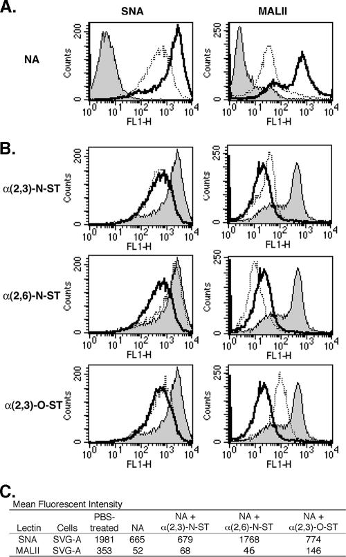
FIG. 1.
Enzymatic treatments of SVG-A cells can deplete and restore cell surface sialic acids. (A) SVG-A cells were treated with 0.25 U/ml NA in PBS buffer (dotted lines) or with PBS alone (bold lines) in suspension at 37°C for 1 h. Cells were incubated with 4 μg/ml biotinylated SNA or 10 μg/ml biotinylated MALII at 4°C for 30 min. To detect lectin binding, the cells were incubated with 3 μg/ml streptavidin-labeled AlexaFluor-488 at 4°C for 30 min. Fluorescence intensity was evaluated using flow cytometry. The shaded histograms represent PBS-treated cells incubated with streptavidin-labeled AlexaFluor-488 only. (B) SVG-A cells were treated with PBS (shaded histograms), 0.25 U/ml NA and then 1 mM NeuAc-CMP (bold lines), or NA followed by NeuAc-CMP and one of the ST enzymes (dotted lines) as follows: 75 mU/ml α(2,3)-N-ST (top row), 25 mU/ml α(2,6)-N-ST (middle row), or 100 mU/ml α(2,3)-O-ST (bottom row). The SVG-A cells were washed and then incubated with biotinylated SNA or biotinylated MALII and streptavidin-labeled AlexaFluor-488 as described above. (C) MFI of SVG-A cells with the treatments described above. FL1-H, fluorescence 1 height.

