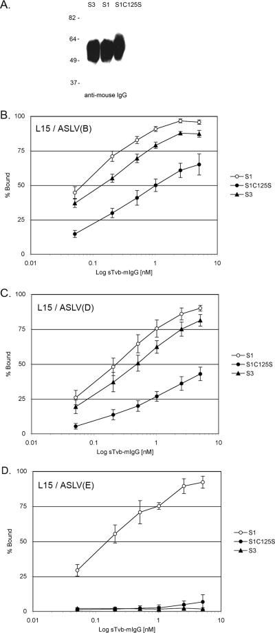
FIG. 3.
Binding affinities of ASLV envelope glycoproteins for Tvb receptors. (A) Western immunoblot analysis of the soluble forms of the Tvb proteins, sTvbS3-mIgG (S3), sTvbS1-mIgG (S1), and sTvbS1C125S (S1C125S), immunoprecipitated with anti-mIgG agarose beads, denatured and separated by sodium dodecyl sulfate-12% polyacrylamide gel electrophoresis, and transferred to nitrocellulose. The mIgG-tagged proteins were probed with anti-mIgG-conjugated to horseradish peroxidase and visualized by chemiluminescence. Molecular masses (in kilodaltons) are given on the left. (B to D) Line L15 CEFs chronically infected with ASLV(B) (B), ASLV(D) (C), or ASLV(E) (D) were fixed with paraformaldehyde and incubated with different amounts of soluble receptor. The viral glycoprotein-soluble-receptor complexes were bound to goat-anti-mIgG linked to phycoerythrin. The amount of phycoerythrin bound to the cells was quantitated by FACS, and the maximum fluorescence was estimated (see Materials and Methods). The data were plotted as percent maximum fluorescence. The values shown are the averages and standard deviations from four experiments.

