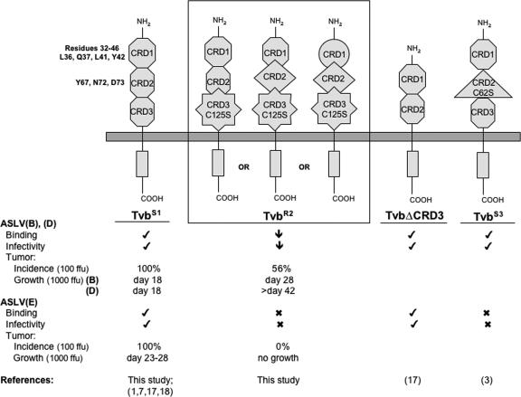
FIG. 5.
Schematic representations of membrane-bound forms of the known and functional chicken Tvb proteins. Hypothetical models are shown for TvbS1, TvbR2 (A, B, and C), the published TvbS1 with CRD3 deleted (TvbΔCDR3), and TvbS3. TvbS1 depicts the wild-type structure of the CRDs (hexagons). The different shapes of the CRDs denote altered structures induced by either the C125S substitution in TvbR2 or the C62S substitution in TvbS3. The abilities of the proteins to function as ASLV(B), ASLV(D), and ASLV(E) receptors are indicated. Check mark, wild-type activity; ↓, significantly reduced activity; X, little or no detectable activity. Tumor incidence (the percentages of chickens infected with 100 FFU of ASVs that formed tumors) and growth (the day when all chickens were sacrificed due to tumor burden from infection with 1,000 FFU ASVs) were summarized from Fig. 4.

