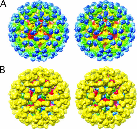
FIG. 5.
Structure of MNV complexed with neutralizing Fab A6.2. (A) Stereo representation of a depth-cued image reconstruction of MNV complexed with Fab fragments from MAb A6.2. Approximately, the constant domains (CH1·CL) of the bound Fab are blue, the variable domains (VH·VL) are green, the protruding domains are green/yellow, and the shell domains are red/orange. (B) Stereo image of the calculated electron density using the modified rNV VLP model and the structure of Fab1 (5), where Fab1 and subunits A through C are yellow, blue, green, and red, respectively.

