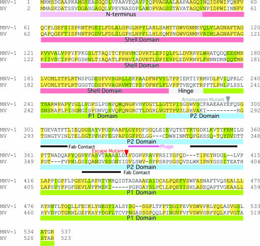
FIG. 7.
NV-versus-MNV-1 sequence alignment. Colored bars beneath the sequences represent the N terminus and the S, hinge, P1, and P2 domains in mauve, red, gray, green, and blue, respectively. The red arrow indicates the location of the known escape mutation (L386F) (23). The black bars indicate the approximate contacts with the antibody, and the purple bar denotes the peptide identified by the phage display that binds to MAb A6.2 (23). The gray arrow denotes the site of the attenuation mutation (36).

