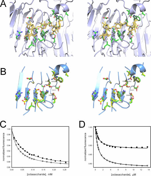
FIG. 5.
O-antigen oligosaccharide binding. (A) Oligosaccharide complex of P22 tail spike protein (pdb entry 1tyx [41]). The octasaccharide, corresponding to two O-antigen repeats from S. enterica serovar Typhimurium, and side chains of amino-acid residues in contact with the ligand are shown as sticks. (B) Binding site residues of P22 (green carbons) and Det7 tail spikes (yellow carbons) aligned by their C-alpha coordinates depicted together with fragments of the Det7 backbone (ribbon diagram). Putative active-site carboxylate residues of Det7 tail spike and the two residues not conserved between both proteins are labeled. (C) Fluorescence titration of Det7tspΔ1-151 with the octasaccharide at 25°C (•) or 10°C (○). The solid lines are nonlinear fits to a binding isotherm for a single class of binding sites resulting in dissociation equilibrium constants of KD, 25°C = 50.7 μM and KD 10°C = 29.9 μM and a 14% fluorescence decrease at saturation. (D) Titration of the P22tspΔ1-108 mutant W365Y (•) and the corresponding wild type (▿) with the octasaccharide at 10°C.

