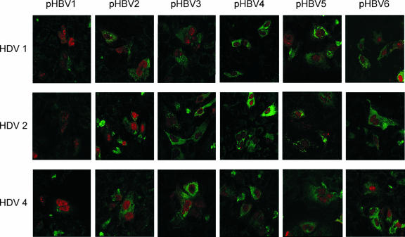
FIG. 5.
Immunofluorescence staining of the expressed HBsAgs and HDAgs in transfected Huh-7 cells examined by using a confocal fluorescence microscope. HDV genotype 1, 2, and 4 expression plasmids (top, middle, and bottom panels, respectively) were cotransfected with six HBV-producing plasmids of genotype B or C. All cells were doubly stained with mouse anti-HBsAg and human anti-HDV antibodies after 3 days of transfection. The secondary antibodies of rhodamine-conjugated rabbit anti-human IgG and FITC-conjugated rabbit anti-mouse IgG were used to show the greenish HBsAg and reddish HDAg immunofluorescence stainings, respectively. Photographs (original magnification, ×630) were taken by using a confocal fluorescence microscope.

