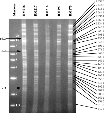
FIG. 7.
Restriction pattern analysis of selected viruses. Virion DNAs from the indicated viruses were double digested with HindIII/NheI, separated by agarose electrophoresis, and stained with ethidium bromide. On the right, the observed restriction fragments from RM175 are correlated with fragment sizes predicted from an assembled sequence file. Arrows on the left indicate fragments of interest that are discussed in the text.

