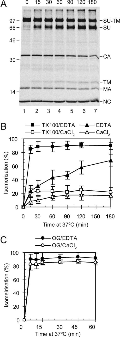
FIG. 1.
In vitro activation of SU-TM disulfide isomerization. [35S]Cys-labeled Mo-MLV was incubated in HN buffer containing either Triton X-100 and EDTA, Triton X-100 and Ca2+, EDTA, OG and EDTA, or OG and Ca2+ for 0 to 180 min at 37°C. A control incubation was done in HN buffer containing Ca2+. NEM was added, all samples were incubated for a total of 180 min, and viral proteins were analyzed by nonreducing SDS-PAGE. Shown are a phosphorimage of a gel with samples incubated in HN buffer with Triton X-100 and EDTA (A) and quantifications of the isomerization efficiencies under all conditions tested (B and C). In the gel analyses SU-TM complexes and free SU and TM subunits are indicated together with other viral proteins to the right and molecular weight standards to the left. The isomerization efficiencies (±standard deviations; n = 6) are given as percentages of complete isomerization.

