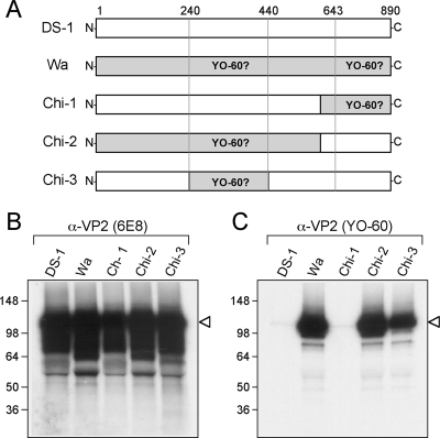
FIG. 7.
Identification of the YO-60-binding region within Wa VP2. (A) Schematic diagram of Wa VP2 chimeric mutants. The diagram illustrates the engineered DS-1/Wa chimeric VP2 proteins. Amino acid numbers (corresponding to Wa VP2) are listed above the proteins. Regions of the protein predicted to be required YO-60 binding are labeled. The chimeric mutants were expressed using rabbit reticulocyte lysate in the presence of [35S]methionine and immunoprecipitated using either 6E8 (B) or YO-60 (C). Proteins were analyzed after SDS-PAGE and fluorography. The images were made from a 12-h exposure of the gels to film. Molecular mass markers (in kilodaltons) are shown on the left of the gels, and the location of VP2 is indicated with an arrowhead.

