Abstract
Paramyxoviruses belong to the Paramyxoviridae family of the order Mononegavirales. They have a nonsegmented negative-stranded RNA genome and can cause a number of diseases in humans and animals. We generated a recombinant Newcastle disease virus (NDV) possessing a two-segmented genome. Each genomic segment is flanked by authentic NDV 3′ and 5′ noncoding termini allowing for efficient replication and transcription. A reporter gene encoding green fluorescent protein (GFP) was inserted into one segment, and a red fluorescent protein dsRed gene was inserted into the other segment in order to easily detect the replication and transcription of segments in infected cells. The rescued viruses grew well and were stable in embryonated chicken eggs over multiple passages. We were able to detect the expression of both reporter genes in the same cell infected with the virus possessing a segmented genome, and viral particles can contain either one or two types of RNA segments. We also rescued a two-segmented virus expressing GFP and the severe acute respiratory syndrome-associated coronavirus spike S protein, which is about 200 kDa. The chimeric virus extends the coding capacity of NDV by 30%, suggesting that the two-segmented NDV can be used for development of vaccines or gene therapy vectors carrying long and multiple transgenes.
The contagious Newcastle disease in birds is caused by Newcastle disease virus (NDV), a single-stranded, negative-sense RNA virus that belongs to the genus Avulavirus of the family Paramyxoviridae in the order Mononegavirales (1). The genomic organization of NDV is similar to that of other paramyxoviruses, containing six transcriptional units that encode the nucleocapsid protein (NP), phosphoprotein and V protein (P/V), matrix (M) protein, fusion (F) protein, hemagglutinin-neuraminidase (HN), and large polymerase (L) protein (16). Each transcription unit contains a conserved transcription start sequence and a 5′ transcription stop signal. The 3′ leader and 5′ trailer regions of NDV that flank the six transcription units are the cis regulatory elements involved in replication, transcription, and packaging of the RNA genome (16). The 3′ leader sequence serves as the sole promoter for viral transcription, resulting in the phenomenon of transcriptional polarity, in which the genes closer to the 3′ end are transcribed more efficiently than those toward the 5′ end (16).
The development of reverse genetics has enabled the generation of infectious negative-sense RNA viruses, such as influenza virus (9, 11, 19, 23), rabies virus (31), vesicular stomatitis virus (17, 38), measles virus (27), Sendai virus (12, 15), and NDV (26, 30), from cloned cDNA. Rescue of the viruses expressing foreign antigens allows for the possibility of these viruses to be used as live attenuated vaccine vectors. Among them, NDV is a unique candidate vector for vaccine antigen delivery in humans and animals (6, 7, 13, 14, 21, 35). Over the last several years, recombinant chimeric NDV Hitchner B1 (NDV/B1) viruses expressing influenza virus hemagglutinin (HA) (21), simian immunodeficiency virus Gag protein (22), or respiratory syncytial virus fusion glycoprotein (20), were rescued and shown to induce specific cellular and humoral immune responses. Recently, a chimeric NDV/B1 virus expressing the ectodomain of HA glycoprotein of a highly pathogenic avian influenza (HPAI) H7N7 virus was also constructed. This virus could serve as a vaccine candidate possessing dual specificity against both HPAI and Newcastle disease in chickens (24).
Besides its ability to carry foreign antigens for induction of immune responses, NDV is also a candidate for cancer therapy in humans. Although it can cause disease in birds, NDV is nonpathogenic to humans, and the majority of humans also lack preexisting immunity to this virus (1). NDV has been shown to specifically replicate in cancer cells that are defective in antiviral interferon production, causing oncolytic effects through activation of apoptotic pathways (8, 10, 18, 25, 29). By using reverse genetics techniques, the HN and the F proteins of NDV can be modified, and the targeting proteins, such as single-chain antibodies against tumor antigens, can be expressed and incorporated into the virus particles (2, 3). These proteins can target NDV specifically to tumors and deliver cancer therapeutic agents into cancer cells (2, 3). Currently, a variety of NDV strains are being investigated in clinical trials against different types of cancers (5, 37).
Despite the advantages of NDV as a potential vaccine vector and cancer therapeutic agent, the ability to carry multiple or long transgenes is limited by the nature of its nonsegmented genome (4, 33). The longest single gene inserted into the NDV genome is the severe acute respiratory syndrome (SARS) virus spike S gene, which is 3,768 bp (6). Our previous experiments also indicated that for the NDV/B1 strain, the insertion of long (>3-kb) or multiple transgenes into its genome is difficult to achieve, and viruses carrying long transgenes have growth defects (unpublished data). On the other hand, there is a demand for the development of NDV vectors that could carry long or multiple antigens or therapeutic molecules. In this study, in order to possibly overcome size limitations, we divided the NDV/B1 genome into two segments and showed that the virus carrying a segmented genome was successfully rescued and stable over multiple passages. Most importantly, we also rescued a two-segmented NDV/B1 virus expressing green fluorescent protein (GFP) and the large SARS virus spike S protein, which is about 200 kDa in size. Our results indicate that an NDV with a segmented genome is capable of expressing a large foreign antigen. The stable two-segmented NDV vector may be an ideal candidate for future multivalent vaccines or cancer therapeutic agents.
MATERIALS AND METHODS
Cells and viruses.
Vero cells were maintained in Dulbecco's modified Eagle's medium containing 10% fetal bovine serum. Chicken embryo fibroblasts (CEFs), prepared from 10-day-old, specific-pathogen-free embryos (Charles River Laboratories, SPAFAS, Preston, CT), were maintained in Eagle's minimal essential medium with 10% fetal bovine serum. Viruses were grown in 8- or 10-day-old embryonated chicken eggs. rNDV/F3aa virus was described previously (24); rNDV/F3aa-GFP virus was rescued from rNDV-GFP cDNA after changing the F-protein cleavage site to a multibasic sequence.
Generation of rNDVs possessing a two-segmented genome.
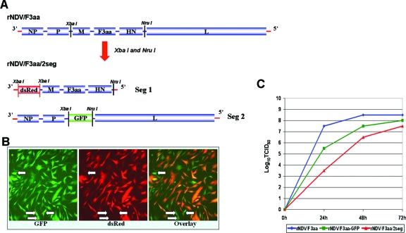
To construct a rNDV/F3aa/2seg virus possessing two genomic segments, the nonsegmented rNDV/F3aa cDNA, which was described before (24), was divided into two parts using two unique restriction sites, XbaI (nucleotides 3163 to 3168) and NruI (nucleotides 8363 to 8368) (Fig. 1A). NruI was generated in this study by site-directed mutagenesis. Both fragments are flanked by authentic 3′ leader and 5′ trailer sequences. A reporter gene dsRed (Clontech) was inserted in front of the M gene of segment 1 (Fig. 1A). A GFP gene amplified from the plasmid phrGFP (Stratagene) was inserted into the XbaI and NruI sites of segment 2 (Fig. 1A). Both dsRed and GFP genes were designed as additional transcriptional units. Recombinant NDV (rNDV) possessing two RNA segments rNDV/F3aa/2seg was then rescued from cDNAs by previously described methods (21, 24). The insertion of new transcriptional units in the recombinant virus was confirmed by reverse transcription-PCR, followed by sequencing. The cDNAs of a two-segmented rNDV/F3aa/2seg-SARS-S virus (see Fig. 3A), which expresses GFP and SARS virus spike S protein (GenBank accession number AY278741), were also constructed using a similar strategy. The virus was rescued again using previously described methods (21, 24). The primers used for cloning and site-directed mutagenesis are available from the authors upon request.
FIG. 1.
Generation of rNDV/F3aa/2seg virus possessing a segmented RNA genome. (A) Construction of cDNAs of rNDV/F3aa/2seg virus. The nonsegmented rNDV/F3aa genome was divided into two segments by using two unique enzyme sites, XbaI and NruI. The authentic 3′ and 5′ noncoding regions were added at the 3′ and 5′ ends of segment 1 (Seg 1) carrying the M, F, and HN genes. The foreign reporter gene dsRed was inserted into segment 1, and the GFP gene was inserted into segment 2 (Seg 2) carrying the NP, P, and L genes. Each segment represents an independent replication unit. (B) Coexpression of GFP and dsRed by rNDV/F3aa/2seg. CEFs were infected with rescued rNDV/F3aa/2seg virus at an MOI of 5. One day after infection, the GFP and dsRed signals were observed using fluorescence microscopy. The arrows designate cells expressing only GFP, not dsRed. (C) Growth kinetics of rNDV/F3aa/2seg in embryonated chicken eggs. Ten-day-old embryonated chicken eggs were inoculated with 100 PFU of each virus, and allantoic fluids were harvested at different time points (24, 48, and 72 h after inoculation). Viral titers (TCID50) were determined in Vero cells by immunofluorescence assay with an anti-NDV rabbit serum and a fluorescein isothiocyanate-conjugated swine anti-rabbit immunoglobulin G (Dako).
FIG. 3.
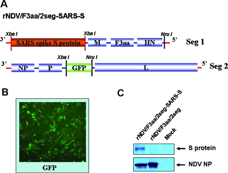
Generation of two-segmented rNDV/F3aa/2seg-SARS-S virus. (A) Construction of cDNAs of rNDV/F3aa/2seg-SARS-S virus. The dsRed gene of the rNDV/F3aa/2seg cDNA segment 1 (Seg 1) in Fig. 1A was replaced with that of the SARS virus spike S gene. The GFP segment 2 (Seg 2) is the same cDNA as used for the rescue of rNDV/F3aa/2seg virus. (B) Expression of GFP by rNDV/F3aa/2seg-SARS-S virus. Vero cells were infected with the rescued rNDV/F3aa/2seg-SARS-S virus at an MOI of 0.1, and 2 days after infection, the GFP signal was observed using fluorescence microscopy. (C) Expression of the spike S protein by rNDV/F3aa/2seg-SARS-S virus. Vero cells were infected with the rescued rNDV/F3aa/2seg-SARS-S and rNDV/F3aa/2seg viruses at an MOI of 0.1 or mock infected. Three days later, cells were harvested, and the expression of the SARS virus spike S protein and the NDV NP protein was detected by Western blotting using monoclonal antibody against spike S protein and rabbit antiserum against NDV, respectively.
Viral growth kinetics.
Embryonated chicken eggs were inoculated with NDV (100 PFU/egg), and allantoic fluids were harvested at different time points after inoculation. The 50% tissue culture infective dose (TCID50) of the virus was determined by immunofluorescence assay in Vero cells.
Immunofluorescence assay.
For the analysis of viral growth and viral protein expression, confluent Vero cells in 96-well plates were infected with viruses in serial 10-fold dilutions. Cells were cultured for 2 days and fixed with 2.5% formaldehyde plus 0.1% Triton X-100. Fixed cells were incubated with anti-NDV rabbit polyclonal serum, washed, and stained with fluorescein isothiocyanate-conjugated anti-rabbit immunoglobulins (Dako). Viral protein expression was examined by fluorescence microscopy.
MDT.
The mean time to death, or mean death time (MDT), was used to determine the pathogenicity of rNDVs in embryonated chicken eggs. Briefly, five 10-day-old embryonated chicken eggs were infected with serial 10-fold dilutions of viruses. The eggs were incubated at 37°C and monitored every 8 h for 7 days. The time to kill the embryos was recorded. The highest dilution that killed all embryos was determined to be the minimum lethal dose. The MDT was calculated as the mean time for the minimum lethal dose to kill the embryos.
Northern blot.
Viruses were inoculated into 8- or 10-day-old embryonated chicken eggs, and the eggs were incubated at 37°C for 3 days. The allantoic fluids were harvested and centrifuged at 6,000 rpm for 30 min at 4°C. The supernatants were laid on a 30% sucrose cushion and centrifuged at 25,000 rpm for 1.5 h at 4°C using a Beckman SW-28 rotor. The pellets were resuspended with phosphate-buffered saline (PBS), and viral RNA was extracted by using TRIzol reagent (Invitrogen). To quantify the viral RNA segments within the cells, CEFs were infected by viruses at a multiplicity of infection (MOI) of 0.1. Two days later, the cells were harvested, and RNA was isolated using TRIzol reagent. The DNA fragment combining the NDV/B1 3′-end 121-bp sequence and the 5′-end 191-bp sequence was labeled with [32P]dCTP using a random primer label kit (Invitrogen) and used as a hybridization probe. RNA blotting was done using NorthernMax solution according to the manufacturer's instructions (Ambion, Inc.). Briefly, viral RNA was loaded onto a 1% formaldehyde agarose gel for electrophoresis. Then, the RNA was blotted onto a nylon membrane (Invitrogen) and exposed to UV irradiation to fix the RNA to the membrane. The membrane was hybridized with 106 cpm/ml probe in QuikHyb hybridization solution (Stratagene), and then the membrane was washed carefully and exposed to a PhosphorImager (Molecular Dynamics) for autoradiography.
Western blot.
Confluent Vero cells in six-well plates were infected with NDVs diluted in PBS containing 0.35% bovine serum albumin and penicillin-streptavidin (Gibco). Two days after infection, the medium was removed and the cells were washed with PBS once. The cells were then lysed in 2× protein loading buffer (100 mM Tris-HCl [pH 6.8], 4% sodium dodecyl sulfate, 20% glycerol, 5% β-mercaptoethanol, and 0.2% bromophenol blue). The protein lysates were separated on a 10% sodium dodecyl sulfate-polyacrylamide gel and transferred to a nitrocellulose membrane (Whatman, Inc.). The membrane was then probed with mouse monoclonal antibody against SARS virus spike S protein (2B3E5; 1 μg/ml) or rabbit polyclonal antiserum against NDV (1:10,000 dilution).
Nucleotide sequence accession numbers.
The GenBank/EMBL/DDJ accession numbers are EU249348, EU249349, and EU249350 for the rNDV/F3aa/2seg dsRed segment 1, GFP segment 2, and rNDV/F3aa/2seg-SARS-S segment 1, respectively.
RESULTS
Generation of an rNDV possessing a two-segmented genome.
To generate the rNDV/F3aa/2seg virus with a segmented genome, the previously constructed full-length rNDV/F3aa cDNA (24) was divided into two parts using two unique enzyme sites, XbaI and NruI, as shown in Fig. 1A. Segment 1 harbors the M, F, and HN genes, and segment 2 contains the NP, P, and L genes. The authentic 3′ and 5′ noncoding regions were added at the ends of segment 1 allowing for replication and transcription. In addition, to detect replication of each segment easily, a reporter gene, the dsRed gene, was inserted in front of the M gene of segment 1, and another reporter gene, the GFP gene, was constructed as an extra transcriptional unit between the P and L genes of segment 2 (Fig. 1A). For most members of the Paramyxovirinae, including NDV, the genome length must be divisible by six to allow for efficient replication. This is known as the “rule of six” (16). Therefore, the nucleotide length of each segment was adjusted to follow this rule. Both reporter genes constitute independent transcription units. An NDV possessing two RNA segments was then rescued from the recombinant cDNAs using reverse genetics as described previously (21). The insertion of new transcriptional units and the separation of the viral genome were confirmed by reverse transcription-PCR, followed by sequencing analysis. When CEFs were infected with the rescued virus at an MOI of 5, we were able to detect both GFP and dsRed protein expression in infected cells (Fig. 1B). We then compared viral growth rates of the rescued two-segmented rNDV/F3aa/2seg, one-segmented rNDV/F3aa (24), and rNDV/F3aa expressing GFP (rNDV/F3aa-GFP) in embryonated chicken eggs. The results showed that rNDV possessing a two-segmented genome was attenuated and grew slower than the rNDV/F3aa or rNDV/F3aa-GFP virus. The maximal titer of rNDV/F3aa/2seg was 10-fold lower than that of rNDV/F3aa (Fig. 1C). These results show that an NDV possessing a segmented genome was successfully rescued and that at least two foreign transcriptional units, GFP and dsRed genes, can be inserted into the viral genome and expressed in host cells.
Stability and virulence of rNDV possessing a two-segmented RNA genome.
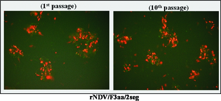
rNDV/F3aa/2seg virus was passaged in 8-day-old embryonated chicken eggs to determine whether the two-segmented virus is stable over multiple passages. As shown in Fig. 2, even after 10 passages, the overall titers were similar, and comparable levels of expression of GFP and dsRed in Vero cells were observed (Fig. 2). The hemagglutination assay (HA) titer of the rNDV/F3aa/2seg virus, which indicates the titer of total particles, correlated well with the infectious titer TCID50 measured in Vero cells at passage 1 and at passage 10. These results suggest that the two-segmented NDV is sufficiently stable and possibly suitable for use as a candidate vaccine vector.
FIG. 2.

Stable expression of GFP and dsRed by rNDV/F3aa/2seg virus over multiple passages. rNDV/F3aa/2seg was passaged in 10-day-old embryonated chicken eggs 10 times. The harvested virus was used to infect Vero cells at an MOI of 0.01. Expression of GFP and dsRed in cells infected with the virus at passage 1 or at passage 10 was observed by using fluorescence microscopy 2 days postinfection.
The virulence of rNDV/F3aa/2seg virus was also compared with the full-length rNDV and rNDV/F3aa viruses in embryonated chicken eggs (Table 1). The MDT was determined after inoculation of each virus into egg embryos. The results showed that the two-segmented virus is significantly attenuated than the full-length viruses. All chicken embryos were alive 7 days after the inoculation.
TABLE 1.
Mean time to death of rNDVs in embryonated chicken eggs
| Virus | Trypsin requirement (cell culture) | Inoculation dose (EID50)a | MDT (h)b |
|---|---|---|---|
| rNDV | Yes | 10 | 108 |
| 1 | 120 | ||
| rNDV/F3aa | No | 10 | 80 |
| 1 | 82 | ||
| rNDV/F3aa/2seg | No | 100 | NA (alive) |
| 10 | NA (alive) | ||
| 1 | NA (alive) |
EID50, 50% egg infective dose.
NA, not applicable.
Generation of two-segmented rNDV/F3aa/2seg-SARS-S virus.
The purpose of dividing the NDV genome into two segments was to increase its capacity to carry long or multiple transgenes. To show this, another two-segmented rNDV/F3aa/2seg-SARS-S virus was rescued. This virus carries two transcriptional units: the GFP gene, which is about 800 bp long, and the SARS virus spike S gene, which is 3,768 bp long and encodes a heavily glycosylated protein of around 200 kDa. The spike S gene was inserted in front of the M gene in segment 1, replacing the dsRed gene (Fig. 3A). The GFP green color was visible in Vero cells infected with rescued rNDV/F3aa/2seg-SARS-S virus (Fig. 3B). In order to show that the rescued two-segmented virus expresses the SARS virus spike S protein, Vero cells were infected with rNDV/F3aa/2seg-SARS-S or rNDV/F3aa/2seg, or they were mock infected. Three days after infection, the cells were harvested, and the lysates were subjected to Western blotting using mouse monoclonal antibody against SARS virus spike S protein or rabbit antiserum against NDV. The 200-kDa S protein was seen only in rNDV/F3aa/2seg-SARS-S-infected cells, not in rNDV/F3aa/2seg-infected or mock-infected cells (Fig. 3C). This virus is also stable over multiple passages.
Rescued two-segmented NDV particles contain either one or two types of RNA segments.
When CEFs were infected with rNDV/F3aa/2seg at an MOI of 5, we detected not only the cells expressing both GFP and dsRed but also the cells that expressed only GFP, as indicated by the arrows (Fig. 1B). For the cells expressing both GFP and dsRed, there are two possibilities. First, the cells may be infected with viruses carrying both genomic segments within one particle: the dsRed segment 1 and the GFP segment 2, thereby expressing both GFP and dsRed proteins. The second possibility is that the cells are coinfected with viruses that carried only one type of RNA segment: segment 1 or segment 2, which could also result in coexpression of GFP and dsRed within one cell.
Interestingly, even at an MOI of 5, we could still detect some cells expressing only GFP and not dsRed (Fig. 1B). The presence of these cells indicates that viral particles carrying only one type of segment, such as the GFP segment, do exist within the viral population. Since the particles carrying only the dsRed RNA segment 1 lack NP, P, and L genes which are required for viral replication and transcription, we could not detect infected cells expressing only the red protein.
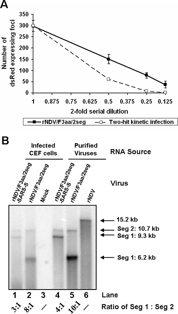
In order to show whether there are viral particles possessing both types of RNA segments (segment 1 and segment 2) within the viral population, CEFs were infected with the rNDV/F3aa/2seg virus in serial twofold dilutions starting at an MOI of less than 0.01. We counted the number of dsRed-expressing foci in each dilution (1, 0.5, 0.25, and 0.125) using fluorescence microscopy. The results showed that the number of dsRed-expressing foci was proportional to the dilutions used for infection. The number of dsRed-expressing foci versus serial dilutions showed linear one-hit kinetics, rather than two-hit kinetics (Fig. 4A). This indicates that the particles containing both types of RNA segments do indeed exist because the number of dsRed-expressing cells decreased linearly in accordance to the dilutions. The expression of dsRed protein requires the presence of both types of RNA segments: segment 1, which provides the dsRed gene, and segment 2, which encodes the RNA-dependent viral polymerase. At an MOI of less than 0.01, there is a very low chance for the cells to be coinfected by more than one particle. Therefore, the cells expressing dsRed must be infected with a single particle possessing both types of RNA segments.
FIG. 4.

Replication-competent two-segmented rNDVs contain both genomic segments in the same particles. (A) CEFs were infected with two-segmented rNDV/F3aa/2seg virus at an MOI of less than 0.01 at serial twofold dilutions. Two days later, the number of foci that expressed dsRed protein was counted in each dilution (1, 0.5, 0.25, and 0.125). The broken line represents the theoretical curve of two-hit kinetics. The solid line represents the actual number of dsRed-expressing foci versus serial dilutions. (B) Ratios of the two genomic RNA segments in two-segmented viral particles or in infected CEFs. RNA was extracted from purified viruses or from CEFs that were either mock infected or infected for 2 days with rNDV/F3aa/2seg or rNDV/F3aa/2seg-SARS-S virus at an MOI of 0.1, run on a 1% formaldehyde agarose gel, and transferred onto a nylon membrane. The membrane was then hybridized with 32P-labeled PCR fragment combining the 3′-end 121-bp sequence and 5′-end 191-bp sequence of the noncoding regions of the NDV genome. The intensities of the bands were quantified by using ImageQuant TL software (Amersham Biosciences), and the ratios of segment 1 (Seg 1) to segment 2 (Seg 2) were calculated for lanes 1, 2, 4, and 5. The RNA purified from nonsegmented rNDV/B1 viral particles was also analyzed, as shown in lane 6.
To determine the efficiency of incorporation of each genomic RNA segment, RNA was purified from rNDV/F3aa/2seg and rNDV/F3aa/2seg-SARS-S virions or from CEFs that were infected with either one of the viruses. The two genomic RNA segments were detected by Northern blotting (Fig. 4B). The precise ratio of the RNA segments to each other was determined by using a hybridization probe: the DNA fragment containing the 3′-end and 5′-end noncoding regions of the NDV/B1 genome. The design of this probe resulted in hybridization to each genomic segment with the same efficiency, allowing determination of the molar ratio of the two segments. Furthermore, to avoid the different transferring rates of the long and short RNA segments during RNA blotting, the Ambion NorthernMax transfer buffer was used, which results in nicking of the longer RNAs. Under these conditions, both the long and short RNAs could be transferred to the membrane at the same efficiency. The molar ratios of segment 1 to segment 2 in purified virions were found to be 10:1 and 4:1 for rNDV/F3aa/2seg and rNDV/F3aa/2seg-SARS-S viruses, respectively (Fig. 4B, lanes 4 and 5). The higher abundance of the shorter segment might result from its higher replication efficiency within the cells. In order to show that this was the case, the ratios of the two genomic RNAs were determined in CEFs infected with the two-segmented NDVs. The molar ratios of segment 1 to segment 2 in infected cells were found to be around 8:1 and 3:1 for rNDV/F3aa/2seg and rNDV/F3aa/2seg-SARS-S viruses, respectively, indicating that the shorter segment indeed replicated more efficiently than the longer one did (Fig. 4B, lanes 1 and 2). Furthermore, in both infected cells and purified virions, the RNA ratios of segment 1 to segment 2 decreased two- to threefold when the dsRed gene was replaced with the long SARS virus spike S gene in segment 1 (Fig. 4B). This indeed demonstrates that the replication efficiency of RNAs of different lengths controls their abundance in purified virions. These results also imply that if the NDV RNA packaging is a random process, then most particles within the two-segmented viral population should contain the shorter segment 1. This further suggests that particles containing only one type of RNA segment exist within the viral population.
DISCUSSION
NDV is a potential vector for generating live attenuated vaccines against various infectious viruses, such as the H5N1 and H7N7 HPAI viruses in birds (13, 24) and SARS virus and human immunodeficiency virus in humans (6, 22). NDV also has intrinsic oncolytic activity and is currently being investigated as a candidate cancer therapy agent (8, 10, 18, 25, 29, 36). In order to deliver different immunogenic antigens to the host or to augment the oncolytic effects by utilizing tumor-targeting molecules, the genome of NDV is required to carry multiple or long transgenes. However, because of the nonsegmented nature and the size of the genome, insertion of foreign genes into the NDV genome is limited. The longer the insert, the more attenuated the chimeric virus is, and the more difficult it is to rescue (4). In this study, we divided the NDV genome into two parts and successfully rescued it (Fig. 1). Since each segment is significantly shorter than the full genome, the virus is able to carry long or multiple insertions. Consequently, we were able to insert the long SARS virus spike S and GFP genes together into the two-segmented viral genome, and the virus expresses both GFP and S proteins in Vero cells (Fig. 3). The total length of the two transgenes in rNDV/F3aa/2seg-SARS-S virus is about 4.6 kb, which is a 30% increase over the original genome. Unpublished evidence from our laboratory indicates that the transgenic loading capacity of the two-segmented NDV can be even higher. Recently, we also rescued a two-segmented NDV which carries three transgenes. In this virus, the SARS virus spike S gene was inserted into the NruI site (after HN) of segment 1 of rNDV/F3aa/2seg virus, while the positions of the other two transgenes, the dsRed and GFP genes, were the same as those in rNDV/F3aa/2seg (Fig. 1A). While both dsRed and GFP were expressed, we were unable to detect the expression of the spike S protein by this virus (unpublished data). The total length of foreign sequences in this virus reaches up to 5.3 kb, which constitutes an increase of almost 35% of the original genomic length. Compared with other negative-stranded RNA viruses, paramyxoviruses are indeed more pleomorphic (32). Rager et al. showed that pleomorphic measles virus particles could package at least two independent genomes at no expense to infectivity (28). Previous work done by Takeda et al. also showed that the genome of measles virus can be divided into segments (34). Not only a two-segmented measles virus but also a three-segmented measles virus could be rescued, and they were able to incorporate up to six additional transcriptional units (34). Exactly how many foreign sequences the segmented NDVs are able to carry still needs to be explored.
As shown in Fig. 1C and Table 1, genome segmentation results in significant attenuation of the virus. One reason for viral attenuation is that the segmented genome alters the original transcriptional polarity. For the two-segmented virus, although the mRNA gradient still exists for each genomic segment, the relative concentrations of mRNAs transcribed from the two genomic segments may be quite different from those of the nonsegmented NDV. The alteration of the transcriptional gradient may result in disproportional protein levels and attenuation of the virus. In the viruses generated in our studies, the three genes required for NDV transcription and replication, the NP, P, and L genes, are located in one segment; and the remaining three genes, the M, F, and HN genes, are located in the other segment. This arrangement may not be the best combination for a two-segmented NDV. In order to minimize virus attenuation, alternative separation and arrangement of the genes in each segment will be required. Another reason that the two-segmented virus is attenuated may be that the viral particles can contain either one or two types of genomic segments, as indicated by this study. The single particles possessing only one segment cannot produce progeny viruses by themselves and thus are defective. To be infectious, at least two particles that contain different segments must coinfect the same cells. In addition, viral particles may carry two or more RNA segments. These particles could contain homogeneous or heterogeneous segments, and only those particles that contain heterogeneous segments are replication competent. In this study, we determined the ratio of RNA segments in two-segmented rNDV/F3aa/2seg virus is 10:1, with the shorter one replicating more efficiently. If the packaging of viral RNAs is a random process and size constraints are disregarded, 82.6% of the particles that possess two RNA segments will contain two copies of segment 1, 0.8% will contain two copies of segment 2, and only the remaining 16.6% will contain one copy of each segment. Therefore, only 16.6% of the particles containing two segments will be replication competent. Although the viral particles may be able to harbor three or more segments, their existence still needs to be demonstrated, and the percentages of these viral particles could be very low if the RNA packaging is random. All those factors could contribute to the attenuation of the two-segmented virus.
In conclusion, in this study we generated a two-segmented NDV that is stable over multiple passages. The segmentation of the NDV genome increases its capacity to carry multiple or long transgenes, adding a new approach for developing NDV as a powerful vaccine vector or cancer therapy agent.
Acknowledgments
We thank Luis A. Martinez-Sobrido for providing the mouse monoclonal antibody against SARS virus spike S protein 2B3E5 and Kathryn A. Fraser and Dmitriy Zamarin for comments on the manuscript. Also, we thank Sa Xiao for performing preliminary experiments.
This work was partially supported by the Northeast Biodefense Center (U54AI057158; Ian Lipkin, principal investigator [PI]), an NIH program project grant (1P01AI59443; Ralph Baric, PI), a Cooperative Agreement (U01AI070469; Adolfo Garcia-Sastre, PI), the Medical Research Center program of MOST/KOSEF (R13-2005-022-02002; Man-Seong Park, PI), and the Bill and Melinda Gates Foundation (grant 38648; David Ho, PI).
Footnotes
Published ahead of print on 16 January 2008.
REFERENCES
- 1.Alexander, D. J. 1997. Newcastle disease and other avian Paramyxoviridae infections. Iowa State University Press, Ames.
- 2.Bian, H., P. Fournier, R. Moormann, B. Peeters, and V. Schirrmacher. 2005. Selective gene transfer in vitro to tumor cells via recombinant Newcastle disease virus. Cancer Gene Ther. 12295-303. [DOI] [PubMed] [Google Scholar]
- 3.Bian, H., P. Fournier, B. Peeters, and V. Schirrmacher. 2005. Tumor-targeted gene transfer in vivo via recombinant Newcastle disease virus modified by a bispecific fusion protein. Int. J. Oncol. 27377-384. [PubMed] [Google Scholar]
- 4.Bukreyev, A., M. H. Skiadopoulos, B. R. Murphy, and P. L. Collins. 2006. Nonsegmented negative-strand viruses as vaccine vectors. J. Virol. 8010293-10306. [DOI] [PMC free article] [PubMed] [Google Scholar]
- 5.Csatary, L. K., G. Gosztonyi, J. Szeberenyi, Z. Fabian, V. Liszka, B. Bodey, and C. M. Csatary. 2004. MTH-68/H oncolytic viral treatment in human high-grade gliomas. J. Neurooncol. 6783-93. [DOI] [PubMed] [Google Scholar]
- 6.DiNapoli, J. M., A. Kotelkin, L. Yang, S. Elankumaran, B. R. Murphy, S. K. Samal, P. L. Collins, and A. Bukreyev. 2007. Newcastle disease virus, a host range-restricted virus, as a vaccine vector for intranasal immunization against emerging pathogens. Proc. Natl. Acad. Sci. USA 1049788-9793. [DOI] [PMC free article] [PubMed] [Google Scholar]
- 7.DiNapoli, J. M., L. Yang, A. Suguitan, Jr., S. Elankumaran, D. W. Dorward, B. R. Murphy, S. K. Samal, P. L. Collins, and A. Bukreyev. 2007. Immunization of primates with a Newcastle disease virus-vectored vaccine via the respiratory tract induces a high titer of serum neutralizing antibodies against highly pathogenic avian influenza virus. J. Virol. 8111560-11568. [DOI] [PMC free article] [PubMed] [Google Scholar]
- 8.Elankumaran, S., D. Rockemann, and S. K. Samal. 2006. Newcastle disease virus exerts oncolysis by both intrinsic and extrinsic caspase-dependent pathways of cell death. J. Virol. 807522-7534. [DOI] [PMC free article] [PubMed] [Google Scholar]
- 9.Enami, M., W. Luytjes, M. Krystal, and P. Palese. 1990. Introduction of site-specific mutations into the genome of influenza virus. Proc. Natl. Acad. Sci. USA 873802-3805. [DOI] [PMC free article] [PubMed] [Google Scholar]
- 10.Fábián, Z., C. M. Csatary, J. Szeberenyi, and L. K. Csatary. 2007. p53-independent endoplasmic reticulum stress-mediated cytotoxicity of a Newcastle disease virus strain in tumor cell lines. J. Virol. 812817-2830. [DOI] [PMC free article] [PubMed] [Google Scholar]
- 11.Fodor, E., L. Devenish, O. G. Engelhardt, P. Palese, G. G. Brownlee, and A. Garcia-Sastre. 1999. Rescue of influenza A virus from recombinant DNA. J. Virol. 739679-9682. [DOI] [PMC free article] [PubMed] [Google Scholar]
- 12.Garcin, D., T. Pelet, P. Calain, L. Roux, J. Curran, and D. Kolakofsky. 1995. A highly recombinogenic system for the recovery of infectious Sendai paramyxovirus from cDNA: generation of a novel copy-back nondefective interfering virus. EMBO J. 146087-6094. [DOI] [PMC free article] [PubMed] [Google Scholar]
- 13.Ge, J., G. Deng, Z. Wen, G. Tian, Y. Wang, J. Shi, X. Wang, Y. Li, S. Hu, Y. Jiang, C. Yang, K. Yu, Z. Bu, and H. Chen. 2007. Newcastle disease virus-based live attenuated vaccine completely protects chickens and mice from lethal challenge of homologous and heterologous H5N1 avian influenza viruses. J. Virol. 81150-158. [DOI] [PMC free article] [PubMed] [Google Scholar]
- 14.Huang, Z., S. Elankumaran, A. S. Yunus, and S. K. Samal. 2004. A recombinant Newcastle disease virus (NDV) expressing VP2 protein of infectious bursal disease virus (IBDV) protects against NDV and IBDV. J. Virol. 7810054-10063. [DOI] [PMC free article] [PubMed] [Google Scholar]
- 15.Kato, A., Y. Sakai, T. Shioda, T. Kondo, M. Nakanishi, and Y. Nagai. 1996. Initiation of Sendai virus multiplication from transfected cDNA or RNA with negative or positive sense. Genes Cells 1569-579. [DOI] [PubMed] [Google Scholar]
- 16.Lamb, R. A., and G. D. Parks. 2007. Paramyxoviridae: the viruses and their replication, p. 1449-1496. In D. M. Knipe and P. M. Howley (ed.), Fields virology, 5th ed. Lippincott Williams & Wilkins, Philadelphia, PA.
- 17.Lawson, N. D., E. A. Stillman, M. A. Whitt, and J. K. Rose. 1995. Recombinant vesicular stomatitis viruses from DNA. Proc. Natl. Acad. Sci. USA 924477-4481. [DOI] [PMC free article] [PubMed] [Google Scholar]
- 18.Lorence, R. M., A. L. Pecora, P. P. Major, S. J. Hotte, S. A. Laurie, M. S. Roberts, W. S. Groene, and M. K. Bamat. 2003. Overview of phase I studies of intravenous administration of PV701, an oncolytic virus. Curr. Opin. Mol. Ther. 5618-624. [PubMed] [Google Scholar]
- 19.Luytjes, W., M. Krystal, M. Enami, J. D. Parvin, and P. Palese. 1989. Amplification, expression, and packaging of foreign gene by influenza virus. Cell 591107-1113. [DOI] [PubMed] [Google Scholar]
- 20.Martinez-Sobrido, L., N. Gitiban, A. Fernandez-Sesma, J. Cros, S. E. Mertz, N. A. Jewell, S. Hammond, E. Flano, R. K. Durbin, A. Garcia-Sastre, and J. E. Durbin. 2006. Protection against respiratory syncytial virus by a recombinant Newcastle disease virus vector. J. Virol. 801130-1139. [DOI] [PMC free article] [PubMed] [Google Scholar]
- 21.Nakaya, T., J. Cros, M. S. Park, Y. Nakaya, H. Zheng, A. Sagrera, E. Villar, A. Garcia-Sastre, and P. Palese. 2001. Recombinant Newcastle disease virus as a vaccine vector. J. Virol. 7511868-11873. [DOI] [PMC free article] [PubMed] [Google Scholar]
- 22.Nakaya, Y., T. Nakaya, M. S. Park, J. Cros, J. Imanishi, P. Palese, and A. Garcia-Sastre. 2004. Induction of cellular immune responses to simian immunodeficiency virus Gag by two recombinant negative-strand RNA virus vectors. J. Virol. 789366-9375. [DOI] [PMC free article] [PubMed] [Google Scholar]
- 23.Neumann, G., T. Watanabe, H. Ito, S. Watanabe, H. Goto, P. Gao, M. Hughes, D. R. Perez, R. Donis, E. Hoffmann, G. Hobom, and Y. Kawaoka. 1999. Generation of influenza A viruses entirely from cloned cDNAs. Proc. Natl. Acad. Sci. USA 969345-9350. [DOI] [PMC free article] [PubMed] [Google Scholar]
- 24.Park, M. S., J. Steel, A. Garcia-Sastre, D. Swayne, and P. Palese. 2006. Engineered viral vaccine constructs with dual specificity: avian influenza and Newcastle disease. Proc. Natl. Acad. Sci. USA 1038203-8208. [DOI] [PMC free article] [PubMed] [Google Scholar]
- 25.Pecora, A. L., N. Rizvi, G. I. Cohen, N. J. Meropol, D. Sterman, J. L. Marshall, S. Goldberg, P. Gross, J. D. O'Neil, W. S. Groene, M. S. Roberts, H. Rabin, M. K. Bamat, and R. M. Lorence. 2002. Phase I trial of intravenous administration of PV701, an oncolytic virus, in patients with advanced solid cancers. J. Clin. Oncol. 202251-2266. [DOI] [PubMed] [Google Scholar]
- 26.Peeters, B. P., O. S. de Leeuw, G. Koch, and A. L. Gielkens. 1999. Rescue of Newcastle disease virus from cloned cDNA: evidence that cleavability of the fusion protein is a major determinant for virulence. J. Virol. 735001-5009. [DOI] [PMC free article] [PubMed] [Google Scholar]
- 27.Radecke, F., P. Spielhofer, H. Schneider, K. Kaelin, M. Huber, C. Dotsch, G. Christiansen, and M. A. Billeter. 1995. Rescue of measles viruses from cloned DNA. EMBO J. 145773-5784. [DOI] [PMC free article] [PubMed] [Google Scholar]
- 28.Rager, M., S. Vongpunsawad, W. P. Duprex, and R. Cattaneo. 2002. Polyploid measles virus with hexameric genome length. EMBO J. 212364-2372. [DOI] [PMC free article] [PubMed] [Google Scholar]
- 29.Reichard, K. W., R. M. Lorence, C. J. Cascino, M. E. Peeples, R. J. Walter, M. B. Fernando, H. M. Reyes, and J. A. Greager. 1992. Newcastle disease virus selectively kills human tumor cells. J. Surg. Res. 52448-453. [DOI] [PubMed] [Google Scholar]
- 30.Romer-Oberdorfer, A., E. Mundt, T. Mebatsion, U. J. Buchholz, and T. C. Mettenleiter. 1999. Generation of recombinant lentogenic Newcastle disease virus from cDNA. J. Gen. Virol. 802987-2995. [DOI] [PubMed] [Google Scholar]
- 31.Schnell, M. J., T. Mebatsion, and K. K. Conzelmann. 1994. Infectious rabies viruses from cloned cDNA. EMBO J. 134195-4203. [DOI] [PMC free article] [PubMed] [Google Scholar]
- 32.Simon, E. H. 1972. The distribution and significance of multiploid virus particles. Prog. Med. Virol. 1436-67. [PubMed] [Google Scholar]
- 33.Skiadopoulos, M. H., S. R. Surman, A. P. Durbin, P. L. Collins, and B. R. Murphy. 2000. Long nucleotide insertions between the HN and L protein coding regions of human parainfluenza virus type 3 yield viruses with temperature-sensitive and attenuation phenotypes. Virology 272225-234. [DOI] [PubMed] [Google Scholar]
- 34.Takeda, M., Y. Nakatsu, S. Ohno, F. Seki, M. Tahara, T. Hashiguchi, and Y. Yanagi. 2006. Generation of measles virus with a segmented RNA genome. J. Virol. 804242-4248. [DOI] [PMC free article] [PubMed] [Google Scholar]
- 35.Veits, J., D. Wiesner, W. Fuchs, B. Hoffmann, H. Granzow, E. Starick, E. Mundt, H. Schirrmeier, T. Mebatsion, T. C. Mettenleiter, and A. Romer-Oberdorfer. 2006. Newcastle disease virus expressing H5 hemagglutinin gene protects chickens against Newcastle disease and avian influenza. Proc. Natl. Acad. Sci. USA 1038197-8202. [DOI] [PMC free article] [PubMed] [Google Scholar]
- 36.Vigil, A., M. S. Park, O. Martinez, M. A. Chua, S. Xiao, J. F. Cros, L. Martinez-Sobrido, S. L. Woo, and A. Garcia-Sastre. 2007. Use of reverse genetics to enhance the oncolytic properties of Newcastle disease virus. Cancer Res. 678285-8292. [DOI] [PubMed] [Google Scholar]
- 37.Wagner, S., C. M. Csatary, G. Gosztonyi, H. C. Koch, C. Hartmann, O. Peters, P. Hernaiz-Driever, A. Theallier-Janko, F. Zintl, A. Langler, J. E. Wolff, and L. K. Csatary. 2006. Combined treatment of pediatric high-grade glioma with the oncolytic viral strain MTH-68/H and oral valproic acid. APMIS 114731-743. [DOI] [PubMed] [Google Scholar]
- 38.Whelan, S. P., L. A. Ball, J. N. Barr, and G. T. Wertz. 1995. Efficient recovery of infectious vesicular stomatitis virus entirely from cDNA clones. Proc. Natl. Acad. Sci. USA 928388-8392. [DOI] [PMC free article] [PubMed] [Google Scholar]


