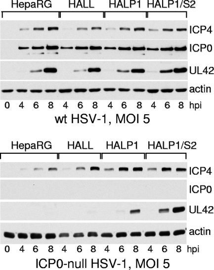
FIG. 7.
Western blot analysis of wt and ICP0-null mutant HSV-1 infection of HepaRG, HALL, HALP1, and HALP1/S2 cells. Cells were infected with wt HSV-1 strain 17 (top) or ICP0-null mutant dl1403 (bottom) at an MOI of 5 PFU per cell in both cases. Samples harvested at the indicated times were analyzed by Western blotting for ICP4, ICP0, and UL42. 0, control mock-infected lanes; hpi, hours post-virus adsorption.

