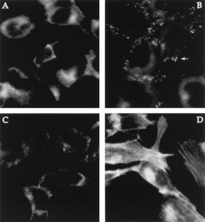
Figure 4.
Formation of focal contacts and stress fibers is induced by carbachol treatment. Immunofluorescence microscopy was used to visualize vinculin (A and B), a major focal adhesion-associated protein, and actin stress fibers (C and D). In serum-starved cells, vinculin staining was diffuse (A), but following exposure to 100 μM carbachol for 10 min, vinculin became concentrated in focal contacts, one of which is indicated by the arrow (B). Similarly, few stress fibers were observed in quiescent cells (C) but were a prominent feature in cells treated with carbachol (D). Actin filaments were detected with a fluorescent phalloidin conjugate. Focal adhesions were visualized with an antibody to vinculin.

