FIG. 2.
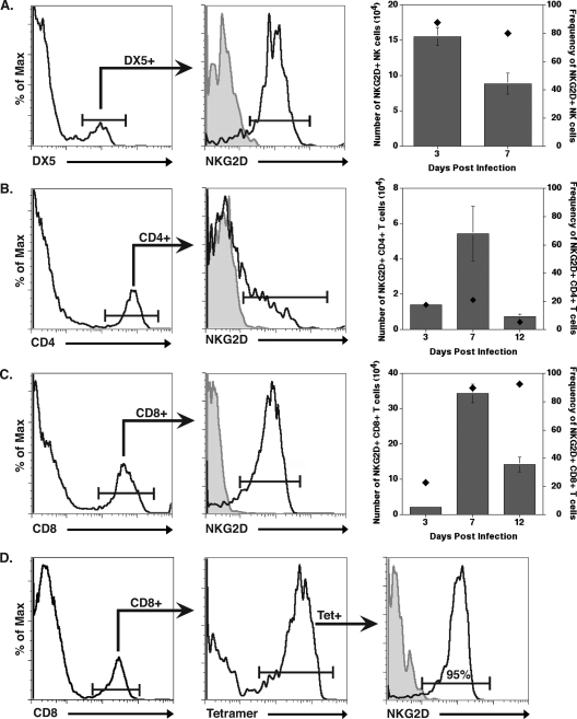
NKG2D expression on cells infiltrating into the CNS of JHMV-infected mice. BALB/c mice were infected i.c. with JHMV, and NKG2D expression on infiltrating cells was determined at select times p.i. NK cells (A), CD4+ T cells (B), and CD8+ T cells (C) were found to express various levels of NKG2D at different times p.i. with virus. Representative histograms (left graphs in panels A to C) are from individual mice at day 7 p.i. For histograms in panels A to C, the shaded area represents isotype-matched control antibody staining and clear areas represent staining with the indicated antibody. In addition, both the overall numbers (gray bars; left y axis) and frequency (black diamonds; right y axis) of NKG2D-positive cells at defined times p.i. are provided (right graphs, panels A to C). (D) NKG2D expression on N318-326 epitope virus-specific CD8+ T cells (determined by tetramer staining) also was defined, with ∼95% of cells at day 12 p.i. expressing NKG2D. Flow cytometric data shown are from a representative experiment of two independent experiments with no fewer than three mice per time point. Data are presented as averages ± SEM.

