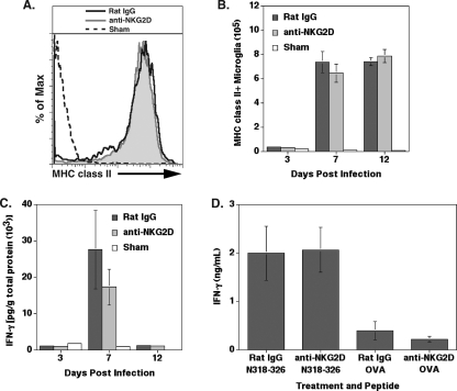
FIG. 5.
NKG2D receptor signaling does not affect IFN-γ secretion in immunocompetent BALB/c mice. IFN-γ secretion within the brain was determined by MHC class II expression on microglia. (A) The fluorescence intensity of MHC class II on the surface of microglia was similar between rat IgG- and anti-NKG2D-treated mice at day 7 p.i., as demonstrated by a representative histogram. (B) Quantification of the mean fluorescence intensity (MFI) revealed that the level of MHC class II on microglia was similar at days 3, 7, and 12 p.i. in mice treated with either anti-NKG2D or control antibody. MHC class II expression data are representative of two independent experiments with no fewer than five mice per group. (C) IFN-γ protein amounts were reduced in the brains of anti-NKG2D-treated mice compared to those of rat IgG-treated mice at day 7 p.i.; however, the difference was not significant. There was no difference in the amounts of IFN-γ protein present in the brain at days 3 and 12 p.i. between treatment groups. Data presented are representative of two separate experiments, with each experiment using a minimum of four mice per time point. Data are presented as averages ± SEM. (D) Anti-NKG2D treatment of total brain isolates obtained at day 7 p.i. and pulsed with CD8+ T-cell epitope N318-326 resulted in increased IFN-γ secretion, but the difference was not significant compared to that of mice treated with rat IgG. Cells incubated with nonspecific OVA peptide are included to illustrate background levels of IFN-γ expression under the experimental conditions used. Data presented are representative of two separate experiments with three mice per experimental condition, and results are presented as averages ± SEM.

