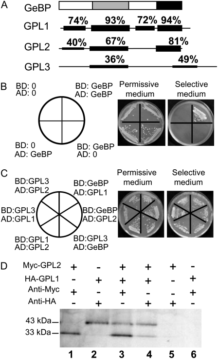
Figure 1.
Dimerization of GeBP and GPL proteins. A, Schematic representation of the GeBP protein and its similarities with GPL proteins. Gray and black areas represent the conserved DNA-binding and C-terminal domains, respectively. B, Homodimerization assay of GeBP in yeast. C, Heterodimerization assay of GeBP and GPL proteins in yeast. D, Coimmunoprecipitation assay of GPL1 and GPL2 proteins in vitro. Myc-tagged GPL2 and HA-tagged GPL1 were translated separately in vitro in the presence of [35S]Met and immunoprecipitated independently with the corresponding anti-tag antibodies. Translation mixes were combined in a 1:1 ratio (lane 3 and lane 4). Immunoprecipitation with the anti-myc antibody (lane 1, lane 3, and lane 6) or with the anti-HA antibody (lane 2, lane 4, and lane 5).

