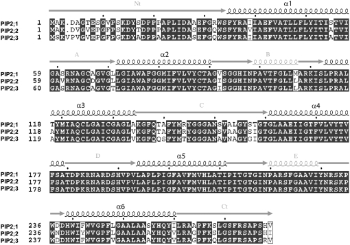
Figure 2.
Alignment of the three encoded proteins PIP2;1, PIP2;2, and PIP2;3. This alignment was made using the programs Mutalin and ESPript (http://nosa-pbil.ibcp.fr). Predicted positions of the transmembrane domains (α1–α6) and external loops (A–E), from N terminus to C terminus, were identical for the three sequences and are shown by the sketches on top of the sequences.

