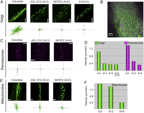
Figure 4.
Roles of myosins XI-2 and XI-K in organelle trafficking in leaves. A, C, and E, Representative images of the indicated organelles (top rows) and paths of individual organelles plotted relative to a common origin (bottom rows; each axis is 100 μm). B, Image of the leaf vein in the Columbia line transformed with the Golgi-specific GFP reporter showing a file of elongated epidermal cells used for organelle tracking. D, Mean velocities of the Golgi stacks and peroxisomes. F, Mean velocities of mitochondria.

