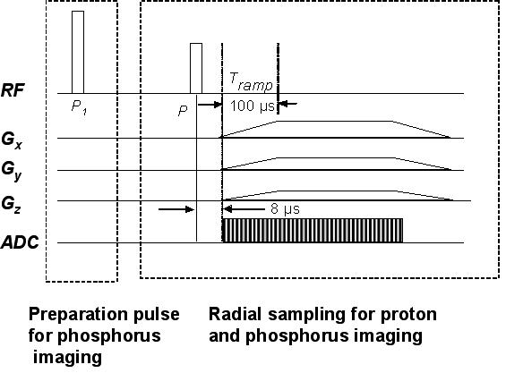
Figure 2.

3D radial projection imaging sequence using ramp sampling for phosphorus and proton imaging designed for uniform mapping of k-space. The initial preparatory pulse, P1 (60°), played out only once, was used for phosphorus with long T1 in order to prepare the magnetization into the steady state.
