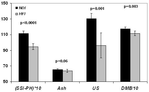
Figure 4 a.

Group differences showing lower measures of bone phosphorus quantified by solid state 31P MRI (SSI-PH) (wt%), ash (wt%), Ultimate strength (US) in N/mm2 and degree of mineralization of bone (DMB) in mg/cm3 in the hypophosphatemic group (HYI) as compared to the same age control group (NOI) in phase I. Bars indicate mean±SD and p represents the statistical significance.
