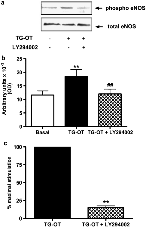
Figure 6.
Involvement of the PI-3-K/AKT pathway in migration induced by TG-oxytocin (TG-OT). (a) HUVECs were preincubated for 30 min with LY294002 (20 μM) before being stimulated with TG-OT (10 nM) for 10 min. Aliquots of cell lysates (30 μg protein per lane) were separated by 7.5% SDS-PAGE and immunoblotted with the indicated antibodies. The experiment was repeated four times with similar results. (b) Densitometric analysis of four independent experiments performed as described in (a). Each bar represents the mean value±s.e.mean. **P<0.001 compared to basal; ##P<0.001 compared to TG-OT. (c) In parallel experiments, HUVECs were pretreated with LY294002 as described in (a) before chemotaxis in the presence of TG-OT (1 nM) as attractant. The results are expressed as the percentage of the maximal migration induced by TG-OT in the absence of LY294002 (100%). Mean values±s.e.mean of four independent experiments. **P<0.001 compared to TG-OT. EGF, epidermal growth factor; HUVECs, human umbilical vein endothelial cells; PI-3-K, phosphatidylinositol-3-kinase; SDS-PAGE, sodium dodecyl sulphate-polyacrylamide gel electrophoresis.

