
Figure 4.
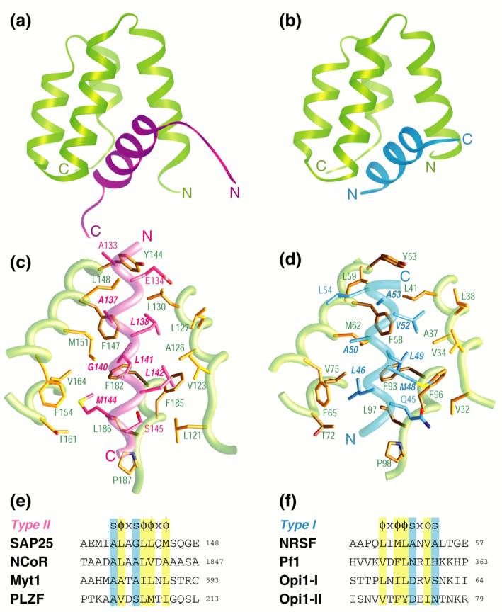
A comparative analysis of intermolecular interactions involving the Sin3 PAH1 domain with diverse targets. A side-by-side comparison of the (a) SAP25 SID-mSin3A PAH1 complex (colored in magenta and green, respectively) and (b) the NRSF SID-mSin3B PAH1 complex (colored in blue and green; PDB code: 2CZY33). Note the contrasting chain directions of the two SID helices. The respective intermolecular interfaces in the two complexes with the backbones rendered as worms and the side chains as sticks (panels (c) and (d)). Residues in the respective SIDs deemed important for the interaction are annotated in bold and italicized. Sequence motifs deduced from structural analysis of the respective complexes (panels (e) and (f); abbreviations: s = any amino acid with a short side chain; x = any non-proline residue; Φ = any bulky hydrophobic residue). The predicted binding sites and modes of a subset of proteins previously shown to target the Sin3 PAH1 domain are also shown. With the exception of yeast Opi1, the proteins are of human or mouse origin (GenBank accession codes: SAP25: NP_001075431; NCoR: AAB17125; Myt1: AAC53456; PLZF: AAD03619; NRSF: NP_005603; Pf1: NP_001028733; Opi1: AAB65073)