
Figure 5. ERα binds to the ERE present in the hLRH-1 promoter.
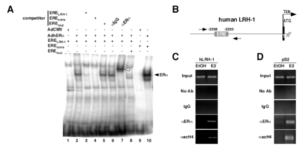
A- EMSA showing binding of ERα to the ERE present in the hLRH-1 promoter. Whole cell nuclear extracts from MDA-MB231 infected with a non recombinant adenovirus (lane 1 and 9) or an adenovirus encoding the human ERα (lane 2 to 8, 10) were incubated with radiolabeled ERELRH-1 (lane 1 to 7), EREmut (lane 8) or EREcons (positive control, lane 9 and 10), in the presence or absence of wild-type (competitor ERELRH-1, lane 3), consensus (competitor EREcons, lane 4) or mutated (competitor EREmut, lane 5) cold probe. Incubation of an anti-ERα antibody resulted in a supershifted band (lane 7, white arrow), whereas no modification in ERα binding was observed with IgG (lane 6).
B- Schematic representation of the 5′ region of the human LRH-1 gene. The ERELRH-1 is indicated. Transcription (arrow, TXN) and translation (ATG) initiation sites are shown. Amplimers are highlighted by the arrows.
C–D ChIP assay demonstrating binding of ERα to the human LRH-1 and pS2 promoters. Cross-linked chromatin from MCF7 treated with the vehicle (EtOH) or E2 10−8M (E2) for 48 hours were incubated without antibody (No Ab), IgG or antibodies against human ERα (aERa) and acetylated histone H4 (aacH4). Immunoprecipitates were analyzed by PCR and positive controls (Input) are shown.