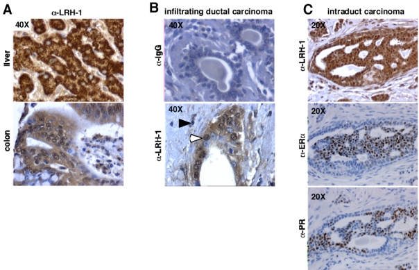
Figure 7. LRH-1 is expressed in tumor cells of mammary infiltrating ductal carcinomas.

A- We used 5 μm formalin-fixed paraffin-embedded liver and colon tissue sections, as a positive control for immunostaining. A strong immunoreactivity was detected on these tissues.
B- As negative controls, mouse IgGs were used instead of the primary antibody on breast tissue section. No staining was observed in these conditions.
C- LRH-1 immunostaining (open arrowheads) is detected in tumor cells of mammary ductal carcinomas, with a nuclear and cytoplasmatic localization. Inflammatory stromal regions surrounding carcinomas contain some lymphocytes positively stained for LRH-1 (filled arrowheads).
