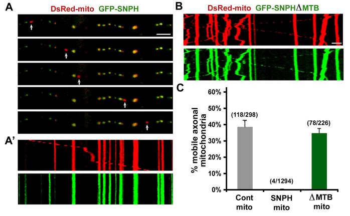
Figure 2. SNPH Immobilizes Axonal Mitochondria.
Neurons were co-transfected at DIV6 with DsRed-mito and GFP-SNPH (A) or GFP-SNPH-ΔMTB mutant lacking the microtubule-binding (MTB) domain (B). Axonal mitochondrial motility was observed in live neurons one week after transfection.
(A) While GFP-SNPH-negative mitochondrion (red, pointed by arrows) migrates along the axonal process, GFP-SNPH-labeled mitochondria (yellow) remain stationary during the time-lapse observation (16 min). (A'), Motion data in (A) is presented in kymograph, in which vertical lines represent stationary mitochondria and slant line or curve indicate motile one.
(B) Kymograph showing the motility of axonal mitochondria labeled with GFP-SNPH-ΔMTB. Scale bars in all panels, 10 μm.
(C) Relative motility of the axonal mitochondria labeled with DsRed-mito alone as controls (n=298 from 16 axons) or co-labeled with DsRed-mito and GFP-SNPH (n=1294 from 39 axons) or DsRed-mito and GFP-SNPH-ΔMTB (n=226 from 9 axons). Error bars: s.e.m.

