
Figure 6. Deletion of the snph Gene Results in Synaptic Short-term Facilitation.
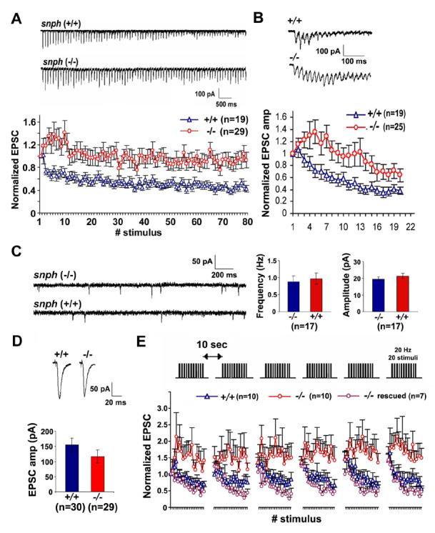
(A, B) Sample traces recorded from the snph (+/+) and (−/−) neurons evoked by 10 Hz, 8 sec (A, upper panel) and 50 Hz, 60 msec (B, upper panel) and their normalized EPSC amplitudes plotted against stimulus number (lower panels of A and B). Substantial short-term facilitation was observed in the snph (−/−) neurons (red circles).
(C) Deletion of the snph gene has no significant effect on the miniature AMPA currents. Left panel: representative miniature AMPA currents recorded from the snph (−/−) and (+/+) hippocampal neurons (DIV14). Right panel: bar graphs of averaged mini AMPA current frequency and amplitude.
(D) Representative EPSCs (upper panels) recorded from paired neurons of the snph (+/+) and (−/−) mice at 0.05 Hz and the bar graph of mean EPSC amplitude (lower panel). No statistical difference was found between the snph (+/+) and (−/−) neurons (P=0.22, t test).
(E) Deletion of the snph gene produces sustained short-term facilitation. 20 Hz, 1 sec stimulus train was delivered repetitively six times at 10-second intervals (upper panel). Normalized EPSC amplitudes were plotted against stimulus number (lower panel). Note that persistent facilitation in synaptic responses was shown only in the snph (−/−) neurons (red circles). Reintroducing the snph gene into the mutant presynaptic neuron (purple circles) eliminates the short-term facilitation and fully rescues the (+/+) phenotype (blue triangles).