
Fig. 1.
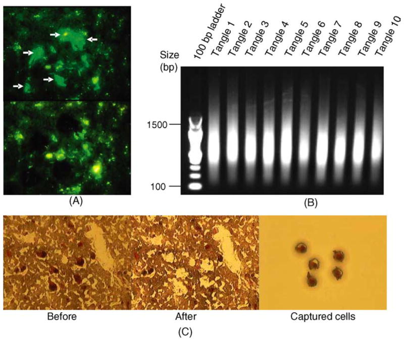
(A) LCM dissected tangle-bearing vs. non-tangle-bearing neurons from the identical AD patient, from the identical brain region. (B) LCM dissected neurons collected from 10 independent AD cases yielded excellent quality RNA following double round amplification and were used to generate expression profiles. Only RNA isolated from tangle-containing neurons is shown. (C) Visualization and isolation of histopathologically normal pyramidal neurons from non-demented control individuals using neutral red staining.