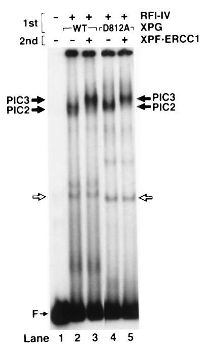
Figure 3.
Detection of preincision complex 3 by electrophoretic mobility shift assay. The substrate was incubated with RFI-IV (XPA, RPA, TFIIH, XPC) and either wild-type or mutant XPG at 30°C to form PIC2 (1st incubation), then XPF⋅ERCC1 was added on ice as indicated (second incubation), and the DNA–protein complexes were analyzed by electrophoretic mobility shift assay.

