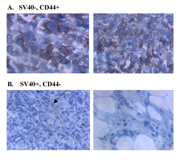
Figure 8.
Examples of human MM in tissue microarrays co-stained using antibodies for SV40 T-antigen (blue/nuclear) and CD44 (red). Of the 34 MMs in the array, approximately 50% were SV40+, and of these SV40+ tumors (~100%) were CD44-. Of the 17 SV40- tumors, approximately 53% were CD44+ (all CD44+ tumors in the tissue array). A. Sample panel of SV40-, CD44+ tumors. B. Sample panel of SV40+, CD44- tumors. The arrow indicates nuclear SV40 T-antigen staining.

