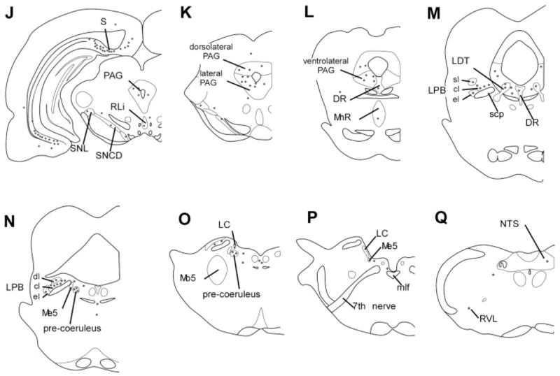
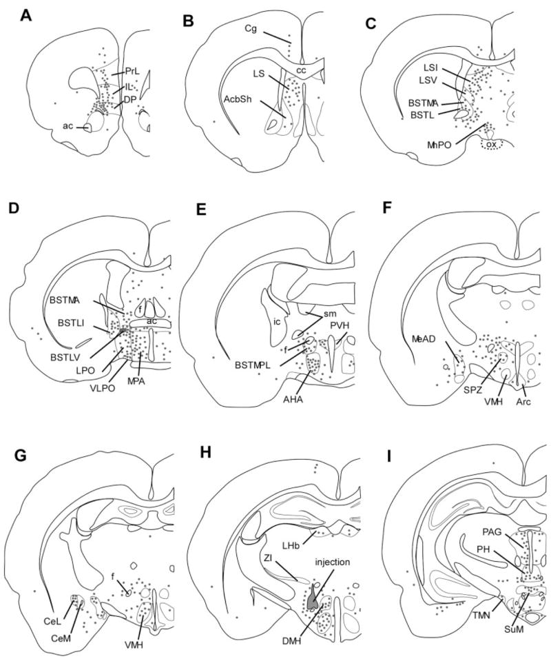
Fig. 3.

Distribution of retrogradely labeled neurons after an injection of CTB into the perifornical part of the orexin field (case 327). Each dot represents five neurons. The heaviest retrograde labeling was in the infralimbic and dorsal peduncular cortices, lateral septum, bed nucleus of the stria terminalis, amygdala, lateral and medial preoptic areas, hypothalamic regions (anterior hypothalamic area, VMH, DMH, PH), and brainstem regions including the PAG, DR, and pre-coeruleus. This injection extended slightly into the zona incerta and also labeled the cingulate cortex, an area not labeled with most other orexin field injections. sl, cl, el, dl, superior lateral, central lateral, external lateral, and dorsal lateral subnuclei of the LPB.

