
Fig. 6.
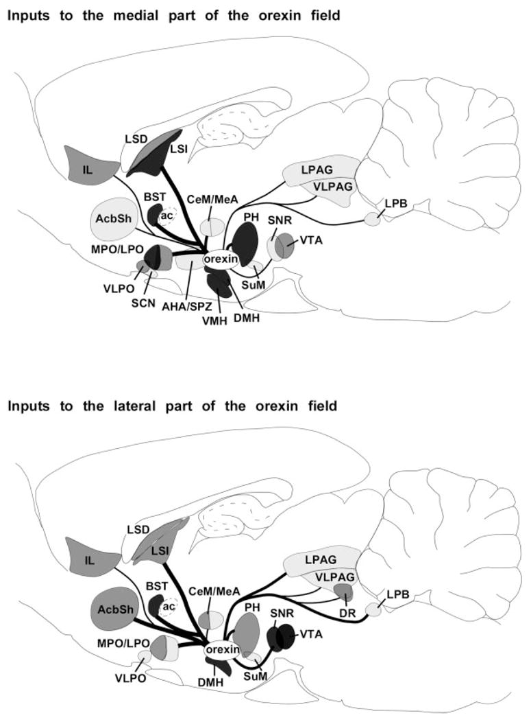
Summary of afferents to the orexin neurons. The upper panel shows inputs to orexin neurons in the medial part of the orexin field and the lower panel shows inputs to orexin neurons in the lateral part of the field. The heaviest inputs are from the pre-optic area, dorsomedial nucleus, posterior hypothalamus, lateral septum, and BST. This pattern suggests that the orexin neurons are influenced by signals regulating emotions, autonomic tone, appetite, circadian rhythms, and sleep/wake behavior. Regions labeled in dark, medium, and light gray innervate >45%, 25–44%, or 5–24% of the orexin neurons. Inputs that innervate <5% of the orexin neurons are not included. Line thickness indicates the relative number of retrogradely labeled neurons.