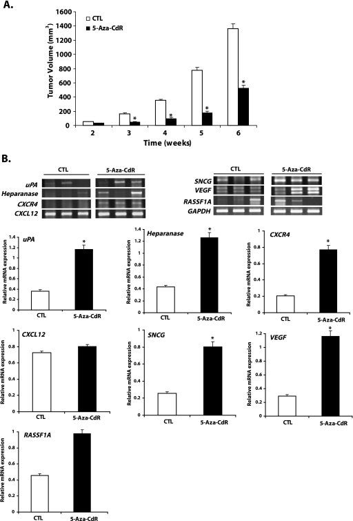
Figure 4.
5-Aza-CdR suppresses tumor growth and upregulates the expression of prometastatic, angiogenetic, and tumor suppressor genes in primary tumor. Female Balb C nu/nu mice were inoculated with either control MCF-7 cells or MCF-7 cells treated with 5 µM 5-aza-CdR for 7 days in the mammary fat pad region in the presence of 0.25 mg of estradiol pellet for a 60-day release. Tumors were measured weekly, and tumor volume was determined as described in the Materials and Methods section. Result represents the mean ± SEM of eight animals in each group. Significant difference from the control is represented by an asterisk (*P < .05) (A). At the termination of the in vivo experiment (week 6), primary tumors were excised and snap-frozen. Total cellular RNA was isolated from these tumors using TRIzol and was analyzed by RT-PCR for the expression of genes responsible for invasion and metastases (uPA, HEPARANASE, CXCR4, CXCL12, and SNCG) and angiogenesis (VEGF) and for the tumor suppressor gene (RASSF1A) as shown (representative three tumor samples from each group) in panel B. The bar diagram represents the mean ± SEM of tumors from three animals in each group (white bars represent mean ± SEM of controls and solid black bars represent mean ± SEM of the 5-aza-CdR-treated group). Significant difference from the control is represented by an asterisk (*P < .05).

