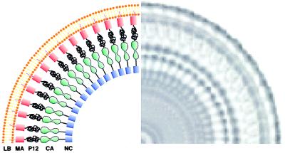
Figure 7.
Schematic model for the packing of the Gag polyprotein in immature murine leukemia virus. The rotationally averaged image of a single particle is shown to the right. The thicker inner leaflet of the lipid bilayer envelope is attributed to the matrix protein (MA), which is known to be anchored to the membrane via a myristoyl group. The low density zone between the MA region and the tracks is assigned to the location of the pp12 protein, which is known to be rich in proline and is likely to be disordered. The ordered track-like structure is assigned to the location of the capsid protein but could also include contributions from the nucleocapsid protein together with bound RNA.

