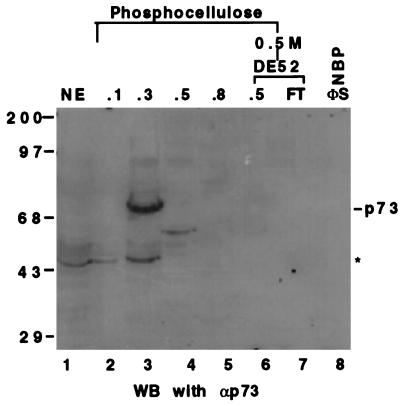
Figure 3.
The Western blot analysis of the fractions from P11 and DE-52 columns using anti-p73 mAbs. Twenty-five microliters of each fraction or HeLa cell nuclear extracts as indicated (Top) was directly loaded onto a SDS/10% polyacrylamide gel. The antibodies recognize both the α and β forms of the p73 protein (35). The asterisk denotes that those signals picked up by the antibodies could be due to nonspecific cross reaction or to the proteolytically degraded products of p73. The band on lane 4 (fraction 0.5 M of P11) may be p73β. The numbers on top indicate the salt concentration that was used to wash the proteins from the columns.

