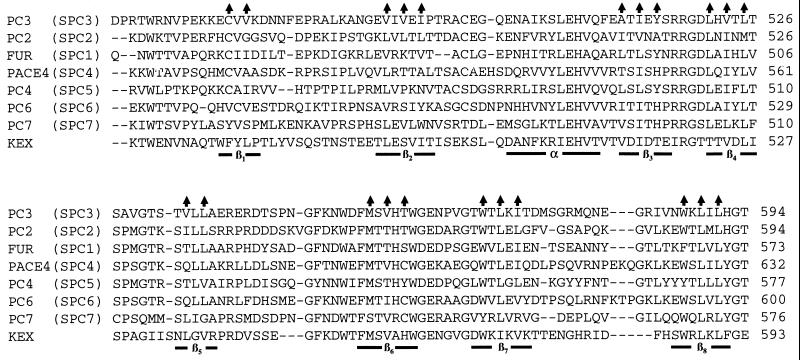
Figure 1.
Amino acid sequence alignment of the P domains of SPC2–SPC7, furin, and kexin (15). The predicted eight β-strands and a region of amphipathic α-helix are underlined. Vertical arrows indicate those β-strand side chains that form the inner hydrophobic core of the P domain.

