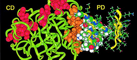
Figure 4.
Conserved framework of the catalytic (CD) and P domains of SPC3 showing proposed dense packing between the hydrophobic surfaces of the catalytic and P domains. The P domain (PD) is shown here in a side view with β-strands oriented vertically. External hydrophobic residues of β-strands 8, 5, and 6 form a hydrophobic patch on the P domain that interacts with the catalytic domain. Surface hydrophobic residues (see Fig. 3B) and conserved Glu and Asp residues of the catalytic groove of the SPCs are shown by orange and red space-filling images, respectively.

