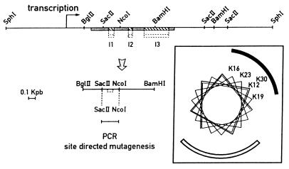
Figure 1.
Restriction map of the genomic clone p3.8Fcos1 containing the psaF gene. psaF exons are shaded and introns are hatched. The strategy used for the site-directed mutagenesis is shown. (Inset) Helical wheel structure of the N terminus of PsaF. The positive and hydrophobic sites are indicated by solid and open curves.

