Figure 3.
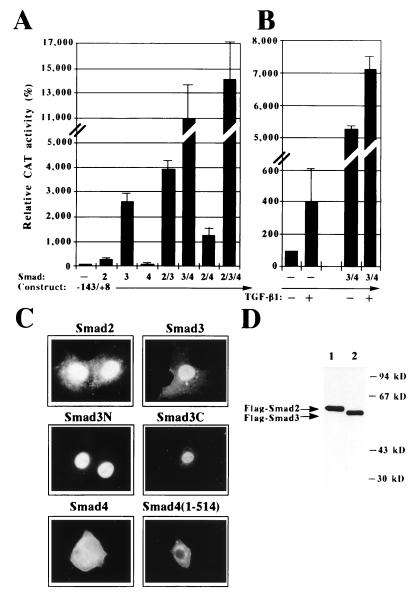
Transactivation of the proximal p21 promoter by Smad family members. (A and B) Transactivation of the −143/+8 p21 promoter by Smad family members. HepG2 cells were cotransfected with the −143/+8 p21 promoter construct in the absence (−) or presence of the indicated Smad proteins. Cells were grown in the absence (A and B, as indicated by −) or in the presence (B, as indicated by +) of TGF-β1. The activity of the −143/+8 p21 promoter in the absence of Smads or TGF-β1 was set arbitrarily to 100%. (C) Subcellular localization of Smad proteins in transfected HepG2 cells. Human Smad proteins were transfected into HepG2 cells and their localization was monitored by indirect immunofluorescence with an antibody against their unique epitope tag [C-terminal FLAG, Smad2 and Smad3; N-terminal FLAG: Smad3C, Smad4, and Smad4(1–514); C-terminal myc, Smad3N]. (D) Western blot analysis of Smad proteins in transfected HepG2 cells. Human Smad proteins 2 (lane 1) and 3 (lane 2) were transfected into HepG2 cells and cell extracts were subjected to SDS/PAGE and Western blot analysis. The resulting chemiluminogram is shown. Arrows indicate the relative migration of the two Smad proteins. Molecular mass markers are in kDa.

