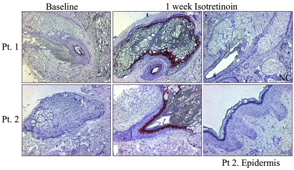
Figure 3. NGAL expression is increased in sebaceous glands in patients biopsied at 1 week of isotretinoin treatment.
Immunohistochemistry using an antibody to NGAL was performed on skin sections taken at baseline and at 1 week of isotretinoin treatment. Sections were incubated overnight with a 1:50 dilution of mouse monoclonal LCN2/NGAL antibody and developed using AEC chromagen (red). All sections were counterstained with hematoxylin. Representative images at baseline and after 1 week isotretinoin from Patients 1 and 2 are shown. An image of the epidermis after isotretinoin treatment from Patient 2 is shown. NGAL was expressed in the sebaceous gland and duct of samples of skin taken at 1 week of isotretinoin therapy. NGAL was not expressed in the epidermis. The amount of NGAL staining varies among patients and individual patient results are shown in Table 4. Negative control consists of normal human skin incubated with normal mouse IgG1 antibody. Original magnification, ×100.

