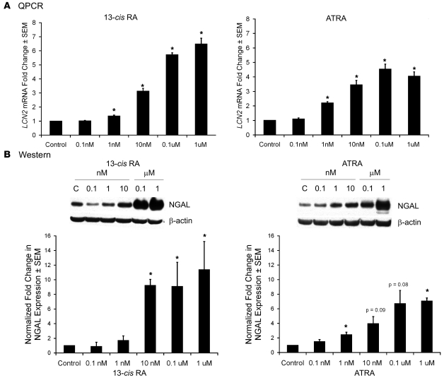
Figure 4. 13-cis RA and ATRA increase LCN2 mRNA and NGAL protein expression in SEB-1 sebocytes.
SEB-1 sebocytes were treated with vehicle as a control (C), 13-cis RA (0.1 nM, 1 nM, 10 nM, 0.1 μM, or 1 μM) or ATRA (0.1 nM, 1 nM, 10 nM, 0.1 μM, or 1 μM) for 48 and 72 hours. (A) LCN2 mRNA expression was verified by QPCR after 48 hours of retinoid treatment. Data represent mean ± SEM of the fold change in gene expression as determined by QPCR of 5 independent samples. Statistical analysis was performed with REST-XL software program and considered significant if *P < 0.05. (B) Protein expression was verified by western blot at 72 hours of retinoid treatment. Blots were incubated with primary antibody to NGAL as well as β-actin as a loading control. Blots were analyzed by densitometry and normalized to β-actin. The graph represents normalized fold-change values (mean ± SEM) relative to control for a minimum of 3 independent blots. Statistical analysis was performed with paired t test; *P < 0.05.

