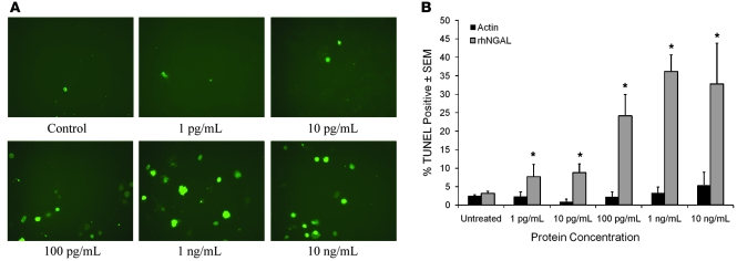
Figure 5. NGAL increases TUNEL staining in SEB-1 sebocytes.
SEB-1 sebocytes were treated with vehicle as a control, 1 pg/ml, 10 pg/ml, 100 pg/ml, 1 ng/ml, or 10 ng/ml of purified rhNGAL protein (R&D Systems) or the same concentrations of human actin protein for 24 hours. (A) Representative images of the TUNEL assay using rhNGAL are shown. Original magnification, ×200. (B) Quantification of the percentage of TUNEL-positive stained cells per treatment at 24 hours. Data represent mean ± SEM; n = 4–8. Parallel experiments to control for nonspecific effects of protein were performed using human actin protein (n = 2). The percentage of TUNEL-positive cells is less than 5% with all concentrations of actin, which is similar to control values. rhNGAL significantly increased TUNEL staining compared to control over a wide range of concentrations with maximal induction noted at 1 ng/ml. Statistical analyses were performed between vehicle control and each treatment concentration of rhNGAL using 2-factor ANOVA with replication; *P < 0.05.

