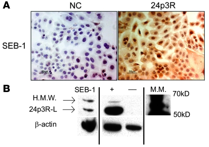
Figure 8. SEB-1 sebocytes express the receptor for NGAL.
(A) SEB-1 sebocytes were grown under standard conditions and immunocytochemistry was performed using an antibody to the murine 24p3R. Slides were counterstained with hematoxylin. Negative control (NC) was processed with normal rabbit IgG antibody in place of the primary antibody. Scale bar: 50 μm. Immunoreactivity for the 24p3R localizes to the cytoplasm of SEB-1 sebocytes. (B) Western analysis confirms presence of the receptor and indicates 2 receptor isoforms are present in SEB-1 sebocytes: high molecular weight (H.M.W.) and 24p3R long (24p3R-L). Positive ([+]; HEK 293 cell lysate) and negative ([–]; T47D cell lysate) controls provided by are shown. All samples were run on the same gel but were noncontiguous. Blot shown is representative of 3 independent experiments.

