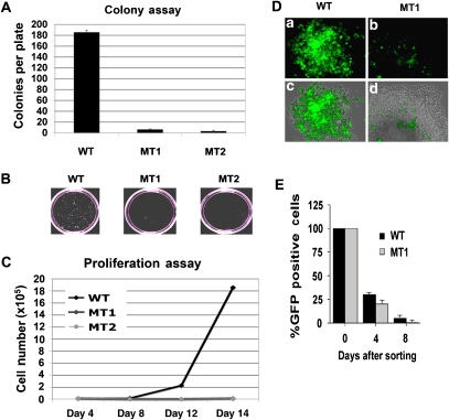
Figure 4.
Host cell growth defects and plasmid instability in KSHV genomes lacking CTCF sites. (A) WT-, MT1-, and MT2-transfected 293 cells were cell sorted for GFP expression and assayed for hygromycin-resistant colony formation. After 10–15 days of selection, the plates were photographed with a 35 mm camera and the colony number was quantitated using Image-Pro Plus software. (B) Example of colony size and density for WT-, MT1-, and MT2-formed colonies. (C) WT-, MT1-, and MT2-transfected 293 cells were sorted for GFP and then cultured in hygromycin-containing media. Cells were then assayed at 4, 8, 12, and 16 days for proliferation using fluorosphere beads and FACS analysis to count cell number. (D) Plasmid stability was assayed by monitoring GFP expression of hygromycin-resistant cell colonies. Hygromycin-resistant cell colony derived with either WT (a, c) or MT1 (b, d); KSHV bacmid was assayed by fluorescence microscopy for GFP (a, b) or a merge of fluorescence with phase-contrast microscopy (c, d). (E) Plasmid stability was assayed by FACS analysis of GFP-positive cells. Identical numbers of WT- and MT1-transfected 293 cells were sorted 4 days after transfection. The percentage of cells retaining GFP-positive signals was then determined 4 or 8 days after initial sorting.

