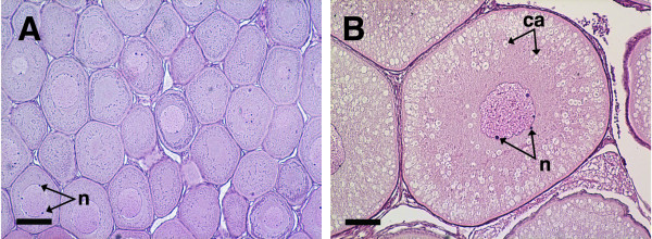
Figure 1.
Histological sections of coho salmon ovaries with perinucleolus (A) and mid-cortical alveolus stage follicles (B). Panel A shows a representative ovary from cohort 1 fish and panel B shows a representative ovary from cohort 2 fish used for subtractive hybridization and qPCR validations. The scale bar = 100 μm in each panel; n, nucleoli; ca, cortical alveoli.

