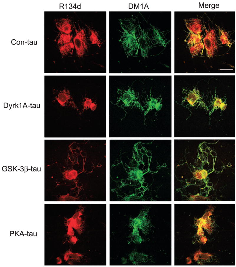
Fig. 4.
Effect of tau phosphorylation at various sites on rebuilding of the microtubule (MT) network in situ. Nocodazole-treated and Triton X-100-extracted 3T3 cells were incubated at 37 °C for 1 h with 15% fresh rat brain cytosol in MT assembly buffer containing 0.5 mg/mL control-treated tau (Con-tau) or tau phosphorylated with dual-specificity tyrosine-phosphorylated and -regulated kinase 1A (Dyrk1A), glycogen synthase kinase-3β (GSK-3β) or cAMP-dependent protein kinase (PKA). The cells were then doubly stained with monoclonal antibody DM1A against tubulin (green) and polyclonal antibody R134d against tau (red), and visualized using a confocal microscope. Bar, 25 μm.

