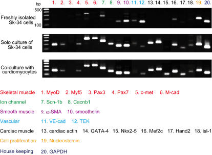
Figure 2. Expression of specific mRNAs for skeletal muscle (1–6), ion channels (7–8), smooth muscle (9–10), vascular markers (11–12), cardiac muscle (13–18) and cell proliferation (19) in freshly isolated cells, and in cells after 5 days of solo culture (without cardiomyocytes) or co-culture with embryonic cardiomyocyte Sk-34 cells.
Note that expression of all 6 cardiomyogenic-specific marker mRNAs can be seen after co-culture with cardiomyocytes. M-cad, M-cadherin; Scn1b, sodium channel voltage gated type1-b; Cacnb1, calcium channel voltage-dependent beta-1 subunit; α-SMA, α-smooth muscle actin; VE-cad, VE-cadherin; TEK, tyrosine kinase-endothelial; GAPDH, glyceraldehydes-3-phosphate dehydrogenase.

