
Figure 2.
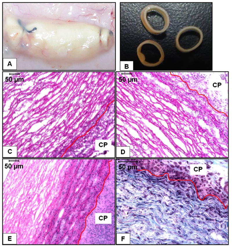
Macroscopic and histological features of explanted EL scaffolds (A) shows the thin capsule over the explants (B) shows the structural integrity of the explants. 7d EL-Gel (C), 7d EL-Gel-FGF (D) and 28d EL-Gel-FGF (E) representative images. Host cells could only infiltrate scaffolds by abluminal side. There is visibly reduced cell infiltration in EL-Gel group. Gomori’s trichrome staining (F) shows new collagen fibers between layers of elastin only in the 28d EL-Gel-FGF group. The red lines indicate the scaffold-capsule interface. H&E stain (cell nuclei are dark blue and matrix is pink), Gomori’s trichrome stain (cell nuclei are dark blue, collagen is blue – green and elastin is pink. Original magnifications (C –E) 200 X and (F) 400 X.