
Figure 3.
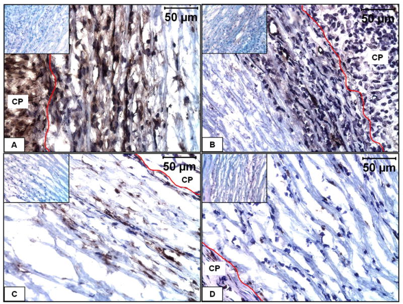
Immunohistochemical characterization of infiltrating cells in EL-Gel-FGF scaffold. A) immunostained for fibroblasts at 28 day, B) immunostained for smooth muscle α-actin at 28 day, C) immunostaining for macrophages at 7 day, D) immunostaining for macrophages at 28 day. The red lines indicate the scaffold-capsule interface. Insets show negative stains. Original magnifications 400 X.