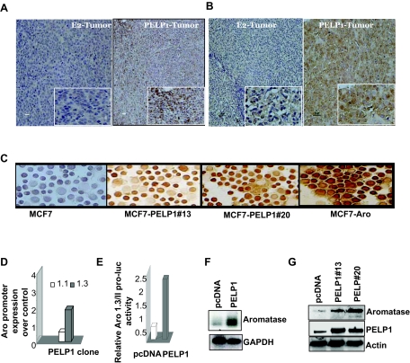
Figure 1.
PELP1 Deregulation Promotes Aromatase Expression
A and B, Immunohistological examination of aromatase expression in mouse xenograft tumor sections from E2-induced or PELP1-induced tumors: A, sections stained with Serotec mAb; B, sections stained with a Novartis mAb. C, Cytospin slides were prepared from control MCF7 and MCF7-PELP1 clones (13 or 20), and expression of aromatase was analyzed by immunohistochemistry. MCF7 Aro cells were used as a positive control. D, Total RNA was isolated from MCF7 and MCF7 PELP1 clone 13 cells and real-time PCR analysis of aromatase gene expression was performed using exon 1.3- and 1.1-specific primers. E, MCF7 and MCF7-PELP1 clone 13 cells were transiently transfected with Aro-1.3/II luciferase reporter, and 48 h later, reporter activity was analyzed. F, Total RNA was extracted from MCF7 and MCF7-PELP1 clone 13 cells, and expression of aromatase RNA was analyzed by RT-PCR. G, Total lysates from MCF7, MCF7-PELP clone 13, and MCF7-PELP1 clone 20 cells were analyzed for aromatase expression using Western blot analysis.

