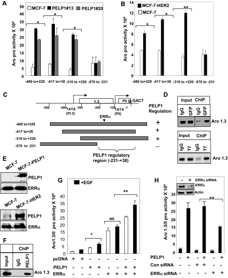
Figure 5.
PELP1/MNAR Is Recruited to the Aromatase Chromatin and Interacts with ERRα
A, MCF7 and MCF7-PELP1 clone cells were transiently transfected with various deletions of Aro-1.3/II luciferase and β-Gal reporter gene and 48 h later treated with 100 ng/ml EGF for 12 h. The reporter gene activation was measured. B, MCF7 and MCF7-HER2 clones were transiently transfected with various deletions of Aro-1.3/II luciferase and β-Gal reporter gene along with PELP1, and 48 h later, reporter gene activation was measured. Data shown are the means ± se from three independent experiments performed in triplicate wells. *, P < 0.05; **, P < 0.001. C, Schematic representation of aromatase deletion constructs used. D, MCF7 cells expressing GFP-PELP1 (upper panel) or T7-PELP1 (bottom panel) were treated with 100 ng/ml EGF for 1 h, and ChIP was performed with IgG, GFP, or T7-tagged antibody. PELP1 recruitment to the aromatase chromatin was analyzed by primers spanning the aromatase 1.3/II region. E, MCF7 and MCF7-PELP1 cells were treated with 100 ng/ml EGF, and nuclear extracts were subjected to immunoprecipitation using an ERRα antibody, followed by Western blot analysis using a PELP1 antibody (upper panel). Nuclear extracts from MCF7 and MCF7-HER2 cells were subjected to immunoprecipitation using an ERRα antibody, and PELP1 presence in the immunoprecipitates was analyzed by Western blot analysis (bottom panel). F, MCF7-HER2 cells were cross-linked with formaldehyde, and ChIP was performed with either IgG or PELP1 antibody. PELP1 recruitment to the aromatase chromatin was analyzed by primers spanning the aromatase 1.3/II region. G, MDA-MB-231 cells were transiently transfected with Aro-1.3/II luciferase and β-Gal reporter gene along with ERRα and PELP1, and 48 h later, reporter gene activation was measured. H, MDA-MB-231 cells were transiently transfected either with ERRα siRNA or control siRNA. After 72 h, cells were transfected with Aro-1.3/II luciferase and β-Gal reporter gene along with PELP1, and 48 h later, reporter gene activation was measured. Inset shows Western blot analysis of ERRα in control and siRNA-transfected cells *, P < 0.05; **, P < 0.001; NS, nonsignificant. Data shown are the means ± se from three independent experiments performed in triplicate wells.

